


Sep 28, 2026

Jul 22, 2026
- Share on:


-
 copied
Selecting the correct CPT code for lumbar decompression surgery depends primarily on the surgical approach and method of visualization. Report CPT 62330 for percutaneous image-guided decompression with indirect visualization, CPT 63047 for open lumbar decompression with direct visualization, and CPT 62380 for endoscopic decompression performed under continuous direct visualization.
Lumbar decompression surgery is done to relieve pressure on the spinal cord or spinal nerves in the lower back to relieve or reduce pain and/or numbness, and improve mobility. This compression is caused by conditions such as herniated disks, spinal stenosis, or spondylolisthesis. Decompression surgery increases space in the spinal canal or neural foramen by removing or trimming parts of the vertebrae, ligaments, or disk material, to reduce the pressure on the spinal cord and nerve roots.
There are 3 different approaches used for lumbar decompression surgery. These include:
- A minimally invasive procedure using percutaneous approach with indirect visualization
- An open approach using direct visualization
- An endoscopic approach where instruments are passed through the scope to perform the procedure.
CPT Definitions of Approach and Method of Visualization
When reporting lumbar decompression procedures, approach and method of visualization are primary differentiators in CPT code selection. For purposes of CPT coding, the following definitions of approach and method of visualization apply to spinal procedures. The primary approach and method of visualization define the service, whether another method is incidentally applied. Surgical services are presumed open, unless otherwise specified.
Approach:
- Percutaneous: Image-guided procedures (eg, computer tomography (CT) or fluoroscopy) performed through a surgical incision with indirect visualization of the spine and without the use of any device that allows visualization through a surgical incision
- Endoscopic: Spinal procedures performed with continuous direct visualization of the spine through an endoscope
- Open: Spinal procedures performed with continuous direct visualization of the spine through a surgical opening
Method of Visualization:
- Indirect visualization: Image-guided (eg, CT or fluoroscopy), not light-based visualization
- Direct visualization: Light-based visualization. Can be performed by the naked eye, or with surgical loupe, microscope, or endoscope
Lumbar Decompression CPT Code Descriptions
62330 (previously 0275T): Decompression, percutaneous, with partial removal of the ligamentum flavum, including laminotomy for access, epidurography, and imaging guidance (ie, CT or fluoroscopy), bilateral; one interspace, lumbar:
CPT 62330 is a minimally invasive procedure performed via a percutaneous approach under indirect vision. No device is used for visualization through surgical incision. A small incision is made and a cannula inserted. Ligamentum flavum and small pieces of vertebral lamina are resected, and tissue and bone sculpting are performed under radiological guidance. Closure of the wound is minimal and could be closed with just steri-strips being applied.
You may see this called a percutaneous image-guided lumbar decompression (PILD).
Report 62331 one time for additional interspaces
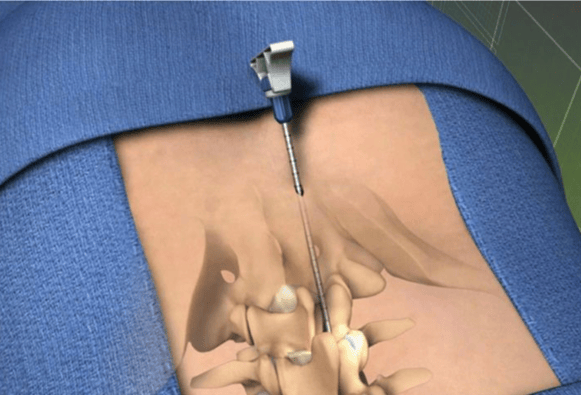
Image courtesy of https://aneskey.com/minimally-invasive-lumbar-decompression-mild-procedure/
63047: Laminectomy, facetectomy and foraminotomy (unilateral or bilateral with decompression of spinal cord, cauda equina and/or nerve root(s), (eg, spinal or lateral recess stenosis), single vertebral segment; lumbar
CPT 63047 uses an open approach with direct visualization of the spine. With the patient prone, a midline incision overlying the affected vertebrae is made. Fascia is incised and paravertebral muscles are retracted. Spinous processes are removed with ronguers. If the stenosis is central, the lamina out to the articular facets are removed using a burr. If the stenosis is in the lateral recess, only half of the lamina is removed. Nerve root canals are freed by additional resection of the facet and compression is relieved by removal of any bony or tissue overgrowth around the foramen.
Report 63048 for each additional vertebral segment
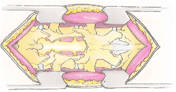
Image courtesy of https://www.researchgate.net/figure/Schematic-lumbar-spinal-stenosis-associated-with-degenerative-spondylolisthesis-and-open_fig1_353536326
62380: Endoscopic decompression of spinal cord, nerve root(s), including laminotomy, partial facetectomy, foraminotomy, discectomy and/or excision of herniated intervertebral disc, 1 interspace, lumbar
CPT 62380 uses an endoscope inserted into a small incision and provides direct visualization of the spine structures and tissues and instruments are passed through the scope to perform the procedure. The endoscope is placed through the foramen and into the spinal canal. Surgical instruments are advanced through the endoscope and herniated disc fragments are removed and the disc is sculpted to eliminate the pressure on the nerve roots.
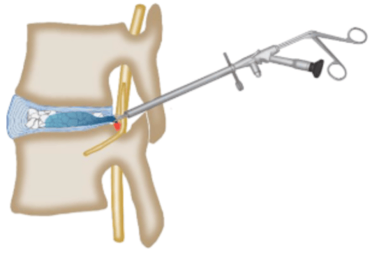
Image courtesy of https://onlinelibrary.wiley.com/doi/full/10.1111/os.12907
Sample Documentation
|
Identify and mark an access point to the interlaminar epidural space of the appropriate level (eg, L3-L4) as close as possible to the midline.... Direct an 18-guage Tuohy needle under fluoroscopic guidance into the epidural space, inject contrast dye to confirm epidural spread.... Make a 1-cm incision at the medial pedicular line approximately 1.5 vertebral levels below the target treatment level. Insert and advance the trocar and portal under fluoroscopic guidance to the interlaminar space.... Advance the trocar to the interlaminar space under fluoroscopic guidance and lock a portal stabilizer device in place. Remove the trocar and attach the depth guide to the portal. Use a small bone rongeur to remove small bony fragments of the superior and inferior laminae under fluoroscopic guidance to allow further advancement of the working portal. Use a tissue sculptor to remove small sections of ligamentum flavum under fluoroscopic guidance with insertion and removal of the tissue sculptor with each resected section....Remove the portal and epidural needle....
63047: Direct visualization with microscope .... Localized the area L4-5 with a spinal needle and fluoroscopy and then it was infiltrated with 1% lidocaine with epi. Incision was made down to the fascia...first dilator was placed and then subsequent dilators were placed and Metrix tube secured. Microscope was then brought in. Used Bovie to dissect and expose the lamina. Drill was then used to create the laminotomy and extended with Kerrison and curette. Ligamentum flavum was identified and removed with Kerrison and thecal sac was seen underneath. Extended out laterally and inferior to expose the lateral edge of the thecal sac. Once satisfied with the decompression retractors were removed....
62380: Endoscopic with direct visualization A needle is inserted through the skin into the disc space via a transforaminal or interlaminar approach.... A skin incision is made, and soft tissue is sequentially dilated over the wire under fluoroscopic control. Special bone burrs or reamers are used to carefully enlarge the foramen under fluoroscopic and guidewire control as much as necessary in order to insert a beveled working tube through which a working-channel endoscope to provide a simultaneous suction and irrigation, as well as to provide bright and clear visualization.... All surgical steps are performed under full, direct visual endoscopic control. All neural structures at L4-L5 are decompressed through removal of herniated intervertebral disc and partial facetectomy. Osteophytes or hypertrophied ligaments are excised as needed with endoscopic shaver blades or drills.... Nerve roots are endoscopically inspected to confirm that they are free of any impingement.... |
Spinal decompression surgery is a procedure aimed at relieving pressure on the spinal cord or nerves due to conditions such as herniated discs or spinal stenosis. It aims to alleviate pain, improve mobility, and enhance the quality of life for patients suffering from spinal conditions. Coding these procedures can be difficult. When making code selections, the main thing to consider is what type of approach was used (percutaneous, open or endoscopic) and whether visualization was direct or indirect with image-guidance.
FAQ
References
- CPT Assistant, March 2026, Volume 36, Issue 3, page 15
- CPT Assistant, February 2017, Volume 27, Issue 2, page 12
- CPT Code Manual CPT Copyright © 2025 American Medical Association.
Since 1992, HIA has been a leading provider of compliance audits, coding support services, and clinical documentation audit services for hospitals, ambulatory surgery centers, physician groups, and other healthcare entities. HIA offers PRN support as well as total outsource support.
The information contained in this coding advice is valid at the time of posting. Viewers are encouraged to research subsequent official guidance in the areas associated with the topic as they can change rapidly.
Need to Earn CEUs?
Our coding education platform, HIAlearn.com, offers courses related to this topic and many more. Subscribe today and access more than 230 courses accepted by AHIMA for CEUs, with a selection also accepted by AAPC.

