Aug 15, 2022

What is “lobar” pneumonia?
“Lobar” pneumonia references a form of pneumonia that affects a specific lobe or lobes of the lung. This is a bacterial pneumonia and is most commonly community acquired. Antibiotics are almost always necessary to clear this type of pneumonia. The antibiotic will be chosen based on the causative organism identified or suspected. This type of pneumonia is also referred to as “non-segmental” or “focal non-segmental” pneumonia and is often referred to in CT of the chest to have the appearance of “ground glass opacity.” Presentation is the same as for other types of pneumonia with dyspnea, productive cough, fever/chills, malaise, pleuritic chest pain and hemoptysis as the common clinical presentation. Complications can include pleural/parapneumonic effusion and empyema. Lobar pneumonia documented by the provider is coded to J18.1 unless the causal organism is specified. Be cautious when using some encoders, as some are still leading the coder to report J18.1 when only the lobe or multilobar is documented. In the ICD-10-CM Alphabetic Index, the coder is referred to see pneumonia, by type. As of October 1, 2019, if pneumonia is documented as affecting a particular lobe, it is coded to J18.9, Pneumonia and NOT J18.1. Lobar pneumonia is a clinical diagnosis made by the physician
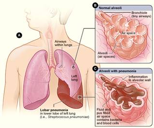
The picture below depicts the lungs and the pneumonia affecting the lower lobe (A). (B) shows normal alveoli and (C) shows infected alveoli.

This type of pneumonia is typically acute with four stages:
- Congestion—within the first 24 hours patient will develop vascular engorgement (the lung becomes heavy and hyperemic)
- Consolidation (red hepatization)—the vascular congestion persists. There is extravasation of red cells in the alveolar spaces. This leads to the appearance of consolidation (solidification) of the alveolar parenchyma
- Grey hepatization—red cells disintegrate. There is still appearance of consolidation but the color is paler and appears drier
- Resolution—complete recovery (exudate will liquefy and will be coughed up in sputum or drain via the lymphatic system
What organism/bacteria is responsible for “lobar” pneumonia?
The most common cause for this type of pneumonia is Streptococcus pneumoniae (pneumococcus). Other common types of bacteria responsible for “lobar” pneumonia are:
- Klebsiella pneumoniae
- Legionella pneumophila
- Haemophilus influenza
- Mycobacterium tuberculosis
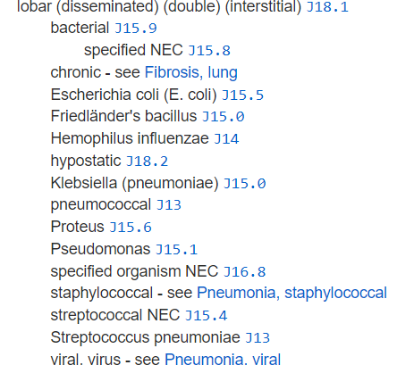
ICD-10-CM Alphabetic Index:
Pneumonia

As you can see above from the ICD-10-CM alphabetic index, if the causative bacteria for the lobar pneumonia is known it would be coded to that specific type of bacterial pneumonia.
Authored by Kim Boy, RHIT, CDIP, CCS, CCS-P
References
https://en.wikipedia.org/wiki/Lobar_pneumonia
https://radiopaedia.org/articles/lobar-pneumonia
https://commons.wikimedia.org/w/index.php?search=lobar+pneumonia&title=Special%3ASearch&go=Go#/media/File:Lobar_pneumonia_illustrated.jpg
AHA coding Clinic for ICD-10-CM/PCS, Third Quarter 2019 Page: 37
AHA Coding Clinic for ICD-10-CM and ICD-10-PCS, Third Quarter 2018 Page 24-25
AHA Coding Clinic for ICD-10-CM and ICD-10-PCS, Third Quarter 2016 Page 15
AHA Coding Clinic Third Quarter 2009 Pages: 16 to 17
AHA Coding Clinic March-April 1985 Page 6
HIA’s comprehensive auditing approach includes acute coding audits and Clinical Documentation Integrity (CDI) audits.
Subscribe to our Newsletter
Recent Blogs
Related blogs from Medical Coding Tips
CMS Hospital Compare is a public reporting pl...
Premier benchmarking is widely used by hospit...
PEPPER reports help U.S. hospitals identify o...
CMS has released the updates to the ICD-10-PC...
Subscribe
to our Newsletter
Weekly medical coding tips and coding education delivered directly to your inbox.


